Your baby is getting bigger. What It Would Look Like Your baby at 29 weeks is stronger more alert and bigger than ever.

Fetal Ultrasound Images 6 Months Babycenter India
29 weeks pregnant lifestyle.
/babyboyultrasound-7bf2ced4b4794754b67dea974b7ec744.jpg)
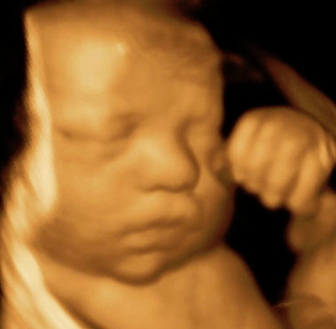
29 weeks pregnant ultrasound. If you take a look at a 29 weeks pregnant ultrasound you will be able to see that the baby is growing white fat deposits under the skin. Your little one measures around 15 inches long from head to heel and hell grow double in size between now and birth. And now the most exciting part Normal 29 week pregnancy ultrasound in case you need one.
2 Ultrasounds Including a 3D by Jen on February 20 2018 I have so much to catch you up on in my 29 weeks pregnancy update. You need to approximately gain 19 to 25 pounds between 20th and 30th week of pregnancy. See how your baby is developing at 29 weeks of pregnancy.
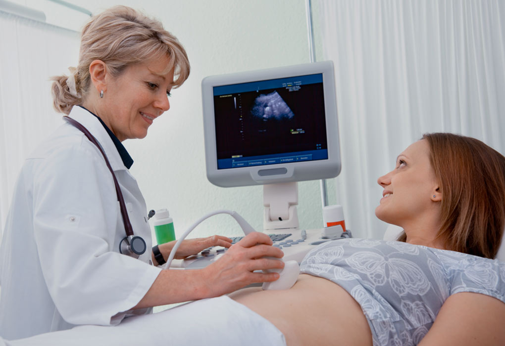
Ultrasound at 29 Weeks Pregnancy Ultrasound in this period is necessary in order to ensure that the state of the baby placenta and the pregnant woman herself is fine. At 29 weeks pregnant most of these will be related to your uterus crowding your digestive system. As the 3rd trimester kicks into gear you are probably noticing a bunch of crazy symptoms.
The most important thing is to eat a healthy and balanced diet. 3D Ultrasound 29 Weeks - YouTube. As your tummy gets heavier its common to have more discomfort.
If youre on a typical prenatal visit schedule you probably dont have to see the doctor at week 29 of pregnancy but youll go back around week 30. If playback doesnt begin shortly try restarting your device. Charlotte Louise Taylor 40550 views.
3D Ultrasound 29 Weeks. If any sign of preterm labor is present such as Contractions Pelvic pressure Backache Cramping or bleeding then an Ultrasound will be ordered. A membrane thin wall forms around your baby.
If you still feel good and dont have any complications you can probably continue your. The fetus size condition of the placenta condition and quantity of amniotic fluid the size and position of the uterus as well as other important information are determined. Your babys skeleton is hardening.
Baby development at 29 weeks. Weve partnered with the American Institute of Ultrasound Medicine AIUM Johns Hopkins and the March of Dimes to create this unique peak into. If you were to look at a 29 weeks pregnant ultrasound you may see that baby is growing white fat deposits under the skin and their energy is surging because of it.
Most mommies who are 29 weeks pregnant report constipation heartburn and even hemorrhoids. I had a pretty big appointment last week at 28 weeks that included an ultrasound and then today I had an elective 3D ultrasound that was just the coolest thing ever. Take healthy diet and include items such as vegetables fruits bran and cereals.
What to expect at 29 weeks pregnant. Most mommies who are 29 weeks pregnant report constipation heartburn and even hemorrhoids. Hes in the process of fine-tuning right now and as his organs continue to develop you may start to.
29 Weeks Pregnant. At 29 weeks pregnant most of these will be related to your uterus crowding your digestive system. Your babys bones are soaking up lots of calcium as they harden so be sure to drink your milk or find another good source of calcium such as cheese yogurt or enriched orange juice.
29 WEEKS PREGNANT - SYMPTOMS HIGH WHITE BLOOD CELLS 29 WEEK BUMP - PREGNANCY UPDATE - Duration. About 250 milligrams of calcium are deposited in your babys skeleton each day. Thats about the size of a butternut squash.
At 29 weeks a baby is almost 10 12 inches 264 centimeters from the top of their head to the bottom of their buttocks known as the crown-rump length and babys height is about 14 34 inches 375 centimeters from the top of their head to their heel crown-heel length. MaryAnn DePietro Week by Week 0. Although you are only a few weeks into your final trimester you may be starting to feel some of the aches and pains of late pregnancy.
We use cookies to give you the best possible experience on our website. 29 weeks pregnant An ultrasound showed too much amniotic fluid and an enlarged cisterna magna no other - Answered by a verified Health Professional. Heres everything you can expect from an ultrasound at this stage in your pregnancy.
29 Weeks Pregnant Ultrasound Baby Position Movement Pregnancy in 29th Week leads you to gain weight. As the 3rd trimester kicks into gear you are probably noticing a bunch of crazy symptoms. 29 Weeks Pregnancy Update.
Your baby now weighs a little over 12kg 25lb and is about 386cm 152in from head crown to heel.
Now that Im further along and the baby has more fat on her and the facial features are stronger I would like to maybe have another ultrasound done. DH and I debated doing the 4D this center doesnt even offer 3D just 2 and 4.

3d Ultrasound At 34 Weeks Page 1 Line 17qq Com
When should I have my 4D ultrasound.

4d ultrasound 34 weeks. Packages FAQs Gallery Testimonials. 3D and 4D Ultrasounds at 34 weeks justcurious2006. After that time most babies are engaged that is they have already gone down into the pelvis and waiting to come out so they can hardly be seen at all on 3D ultrasound.
10 years on and he still looks like our first sight of the little man. 25- 30 Minutes 3D4DHD Ultrasound 28-34 Weeks Fetal Heart Rate Gender Free Recheck Flash DriveCD of all Sugar Spice Package 50 Each package comes with a printout of your bundle of joy. At this point is it pointless since the baby will be here soon and at this late in my pregnancy will I.
3D 5D ultrasound images and 4D ultrasound video can be obtained at any stage. The 3D4D image at my as looks much different from my 34 week elective. 12 Black and White prints.
Gender determination upon request. Included in the Sweet Dreams Package. - Refreshments and a sweet treat - DVD of your session 20 Value.
Best between 24-34 weeks of pregnancy Each session will be 20-25 minute 3D4D ultrasound session. Listen to your babys heartbeat. FREE CD with unlimited 2D and 3D images.
The latest that we do 3D4D ultrasounds is at 32 weeks. 5 Color Thermal 5 Black and White. 30 weeks 4d ultrasound disappointment.
Experts also discourage the use of any kinds of ultrasounds 2D Doppler 3D and 4D for the purpose of creating a memento. I have made an appointment for June 9th. Hi ladies I had the 4D ultrasound today however bubs face didnt show very much and not clear at all.
34 Week 3D ultrasound. This is will be my first and only ultrasound because its what my MW requires per contract. A 3D4D Ultrasound taken of a baby at 34 weeks that visited Prenatal Impressions the only premier provider of 3D4D Ultrasounds in Orlando Florida since 20.
Surprise Mom or make it a planned event. Many claim the optimum time is somewhere between 27-32 weeks of gestation and others state between 26- 30 weeks. There is some discrepancy between clinics around when a 3D andor 4D ultrasound should be done.
34 Week 4d Ultrasound Results from Microsoft. At this stage the baby has put on some weight and filled out to make features more visible yet still enough fluid in front of babys face to obtain great images. However we do recommend a gestational age of 26-34 weeks for the best facial detail.
FREE 4D live DVD video of your session set to music. Fun family games are optional. 12 4x6 Color Prints.
Yes she had her hand on her face but even when she removed it the pictures are not clear enough and that really disappointed me because I still had to pay and the lady wouldnt keep trying to get a better shot. Currently ACOG recommends that expecting women have at least one 2D ultrasound between weeks 18 to 22 of pregnancy noting that some women may also have a first-trimester ultrasound. 14 Weeks Length.
99 3D4D Ultrasound Special. 4D ultrasound 34 weeks pregnant face 4D Rafael Ortega Muñoz MD Ciudad Real. 4d ultrasound 34 weeks.
This is my husbands 1st child and this is something that I want to give him for his 1st fathers day. You receive everything at your appointment. 4D ultrasound 34 weeks pregnant face 4D Rafael Ortega Muñoz MD Ciudad Real - YouTube.
Enjoy our 3D4D Ultrasound Studio for a full hour with all of your closest friends and family. I had a 3D4D ultrasound done at like 24 weeks. I was wondering how good you can see everything at 34 Weeks on a 3D and 4D scan.
The suggested results are not a substitute for clinical judgment. Ultrasounds done later in the pregnancy are less accurate for dating so if your due date is set in the first trimester it shouldnt be changed.
Crown-rump length of 15-60 mm was superior to BPD but then BPD at least 21 mm was more precise.

Ultrasound due date accuracy. If the ultrasound date is within seven days of your LMP date we would stick with your LMP date. Also what is more accurate ultrasound due date. You mentioned that youre about 24 weeks along you had an early ultrasound when you were 6 weeks and you werent sure when your last period was.
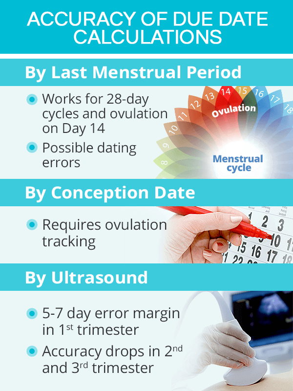
Pregnancy ultrasound does give an idea to the expecting mother about the tentative date of delivery. If there is a discrepancy in your recall of LMP and the ultrasound results most doctors will go with the ultrasound results. Evidence suggests that ultrasounds more accurately predict your due date than using your last menstrual periodbut only in the first trimester and early second trimester until roughly 20 weeks.
Also how some time after the procedure for you and your partner to have a coffee or lunch to chat about the scan and how it went. Enter the Calculated Gestational Age on the Date Ultrasound was Performed. Although I KNOW waiting and being late etc is awful the baby WILL eventually come.
Generally - 1 week in first trimester - 2 weeks in second trimester. Join Huggies now to receive week by. The accuracy of ultrasound varies depending on when in pregnancy it is done.
Ultrasound examinations from 12 to 22 weeks are regarded as being within 10 days of accuracy or up to 10 days earlier or 10 days later than the womans calculated due date. I think it depends on many things. The accuracy of the EDD by ultrasound depends on several factors such as the current stage of pregnancy the quality of the machine and the position of the baby in the mothers womb.
Ultrasounds performed after 22 weeks gestation cannot be used to estimate the due date of the baby because the size no longer reflects the age very well. Ultrasound was more accurate than LMP in dating and when it was used the number of postterm pregnancies decreased. All calculations must be confirmed before use.
Can Your Pregnancy Ultrasound Determine Your Due Date. My due date is about 5 days different from my LMP and my scan datesI am going with my last scan date as that was most accurate with my last daughter. Early ultrasound due dates have a margin of error of roughly 12 weeks.
This is because a semi full bladder will help to push your are up higher in accurate pelvis making it easier for the sonographer to see. Calculate Due Date or Gestational Age Using Dates. The most accurate time is between 8 and 11 weeks gestation.
Combining more than one ultrasonic. Ultrasounds performed during the first 12 weeks of pregnancy are generally within 3 5 days of accuracy. 20w ultrasound due date confusion.
Due Date Accuracy Early ultrasound due dates have a margin of error of roughly 12 weeks so doctors will usually keep the original due date the one generated by the date of your last menstrual period if the ultrasound due date is within that margin of error1. The procedure accurate takes around 45 minutes from start to finish. As mentioned earlier the due date which is calculated from the last menstrual cycle often does not match the due date calculated by ultrasound.
The earlier the ultrasound is done the more accurate it is at estimating the babys due date.
Around 5 weeks the gestational sac is often the first thing that most transvaginal ultrasounds can detect. What Can I See on Ultrasound at 5 Weeks.

Pin On My 3rd Pregnancy 16 Years Later
Monochorionic twins are detected by heart beat after 6 full weeks.

Ultrasound at 5 weeks. This is seen before a recognizable embryo can be seen. Prompt diagnosis made possible by transvaginal ultrasound can. If you get a vaginal scan at 5 weeks the sonographer should be able to able to detect two separate gestational sacs if youre expecting dichorionic twins.
On your 5 weeks pregnant ultrasound you should be able to see your gestational sac and the yolk sac which is always present when you are 5 weeks pregnant. What is the normal size of gestational sac at 5 weeks. Twin ultrasound 5 weeks What can you detect.
If you are carrying fraternal twins you will be able to see the yolk sac as well as the fetal poles in two separate sacs. If youre 5 weeks pregnant your ultrasound will be done via the vagina as opposed to transabdominal ultrasounds that are typically performed later on in pregnancy. Transvaginal ultrasound by contrast can detect pregnancies earlier at approximately 4 ½ to 5 weeks gestation.
Within this time period a yolk sac can be seen inside the gestational sac.
AP 6 week ultrasound twins 8 weeks one baby 12 weeks one baby. Twiniversity is the 1 resource for expecting twins and raising twins including expecting twins classes free articles a free podcast a twin mom mentor program and so much more.
Seeing twins at 6 weeks is definitely possible.

6 week ultrasound twins. Heard our little ones heart beat which I was so relieved to see and hear. Returning to how twins would appear we. Will 6 Week Ultrasound Show If You Have Twins.
We were able to see our baby and see the heart flicker. Videos you watch may be. At about 5-6 weeks a tiny structure termed the fetal pole becomes visible along with a ring-shaped structure called a yolk-sac.
Those tiny pea pods that are barely a centimetre in size will grow to be full-formed babies within no time. I was super emotional and couldnt even th. They missed my twin on my 6 week scan found them 10 days later to check and make sure babys heartbeat was going up.
Twins can be detected on an ultrasound in the first trimester as early as 4-6 weeks after you miss your period and their heartbeats can be found at 6-8 weeks its also pretty hard to distinguish two heartbeats and having two doesnt always indicate twins. Anatomy scan at 20 weeks. Your babies will be developing rapidly but a 6 week ultrasound for twins provides you with more details about your multiples.
Hello all Im new here and just wanted some opinions. At 6 weeks youll likely have a transvaginal ultrasound rather than the abdominal one you may be thinking of. I had an abdominal ultrasound at 6 weeks which showed one baby and i reaaally think its twins because im having the same symptoms i had with my twins.
By four weeks twins will start showing during ultrasound in form of 2 gestational sacs but you cannot get clear indication of twins until 6 weeks. These ultrasounds will also show the heartbeat of the growing fetuses. Boba87 Mon 31-Jul-17 221519.
7 Posts Add message Report. Blighted ovum ultrasound 6 weeks Are 6 week ultrasounds accurate Connect by text or video with a US. Learn the symptoms at 6 weeks and find out what to do next if you are really having twins.
A twin ultrasound at 6 weeks needs to be done vaginally to detect twins this early in your pregnancy. Had my first ultrasound at 65 weeks. The exact time twins can be detected depends on the type of twins for example if theyre identical from one egg or not.
The tech said she was SURE it was just one baby. Hidden twins are more common with early ultrasound because the baby is of course significantly smaller than later in the pregnancy Plus it depends on. They think I may have ovulated twice.
I have another ultrasound on thursday which is abdominal as well and i think it should show twins if there are any. I had asked if it was twins 4 weeks later same tech does the scan and I think she was more shocked than I was that there were 2 babies on that screen. Week 6 Ultrasound 6W5D Twins At 6w5d gestation you can see that each embryo 8 mm each.
6 week ultrasound twins. This is when youll know for sure whether or not your expecting twins. She said in 35 years this is only the second time shes ever seen it happen.
Our early 6 week ultrasound was scheduled due to spotting. Observing twins at 6 weeks on the ultrasound scan is what makes most couples feel like parents for the first time. Twin A measured 6wk 4 day and heartbeat 115 Twin B measured 6wk 1 day and heartbeat 105 As sac is not as big as it appears.
It is the angle on the us. Six full weeks is when youre 60 weeks pregnant. Many parents are curious to know if theyre having twins.
By week 6 or 7 of your pregnancy an ultrasound will definitively reveal that youre pregnant with twins. Just measure that out on a ruler. When youre 60 weeks pregnant your babys hearts should be beating and the sonographer should be able to detect them at this point.
6 week ultrasound - Twins. When youre 6 weeks pregnant with twins you are between 5. At this stage the presence of two yolk sacs can be seen and separate heartbeats distinguished.
Robinson Triplets - 6 week Ultrasound twins If playback doesnt begin shortly try restarting your device. Robinson Triplets - 6 week Ultrasound twins - YouTube. At 6 weeks there should be a visible fetus yolk-sac and the visible flicker of a fetal heartbeat.
I had an early scan today my midwife estimated me to be 6wk6d. Thats also called being 7 weeks pregnant. At an ultrasound scan at 6 full weeks a sonographer will most likely be able to spot any type of twin pregnancy.
Ultrasounds taken at this date make sure that the pregnancy is taking place in the uterus. Before 7 weeks babies are often so small that the abdominal ultrasound may have. How do you know if youre 6 weeks pregnant with TWINS.
Board-certified doctor now wait time is less than 1 minute.
Halaman
Baby & Child
Cari Blog Ini
Label
- 13th
- 14th
- 16th
- 2015
- 2016
- 2017
- abbott
- abortion
- about
- accuracy
- accurate
- ache
- achy
- acid
- acne
- activities
- activity
- acts
- acupuncture
- adhd
- adopt
- adopting
- adoption
- adulthood
- adults
- advanced
- advent
- advice
- after
- agencies
- agency
- alive
- allergic
- allergy
- along
- aloud
- alton
- amazon
- american
- ammonia
- amoxicillin
- anatomically
- andrea
- angel
- anger
- animal
- animals
- animated
- anna
- announcement
- announcements
- announcing
- answers
- anti
- antonio
- anxiety
- applesauce
- apps
- aquaphor
- arms
- arts
- aspirator
- aspirin
- atlanta
- attractions
- autism
- autistic
- average
- away
- babies
- baby
- babys
- babyshower
- babysit
- babysitting
- babywearing
- back
- backpack
- bags
- bahamas
- bake
- baked
- ball
- balloon
- balls
- banana
- band
- barometric
- basal
- base
- baseball
- bassinet
- bath
- bathing
- bathroom
- battle
- bauer
- bday
- beach
- beaches
- bean
- beautiful
- bedding
- beds
- beef
- beets
- before
- behavior
- behind
- being
- bell
- belly
- belt
- benadryl
- bench
- best
- better
- between
- beyond
- bible
- bike
- biography
- birth
- birthday
- bismol
- bite
- bites
- black
- blank
- blanket
- bleach
- bleeding
- blended
- block
- blog
- blood
- bloody
- blue
- blueberry
- board
- boat
- body
- book
- books
- booster
- boot
- boppy
- borax
- born
- bottle
- bottles
- bouncers
- boxes
- boyfriend
- boys
- brace
- bracelets
- bradley
- braids
- brazilian
- break
- breakthrough
- breast
- breastfeeding
- breastmilk
- breath
- breathe
- bright
- brisket
- britax
- broil
- brother
- brown
- brunch
- budget
- build
- bull
- bump
- bumped
- bumps
- bunk
- burping
- busbys
- busters
- butterfly
- buying
- buzz
- cabbage
- caff
- caffeine
- cake
- cakes
- calendar
- california
- call
- calls
- calorie
- camp
- camping
- camps
- cancun
- candy
- canker
- canopy
- cape
- capsule
- caput
- cardboard
- care
- carey
- carolina
- carolinas
- carpal
- carrier
- carriers
- carrot
- cars
- carseat
- casserole
- cast
- castaway
- casting
- catering
- cause
- cedars
- ceiling
- center
- centerpiece
- centerpieces
- cereal
- certificate
- cervix
- chair
- chairs
- challenge
- chamomile
- chance
- chances
- change
- changing
- chapter
- character
- chart
- cheap
- cheating
- check
- cheese
- cheesecake
- chef
- chest
- chicago
- chicco
- chicken
- child
- childbirth
- childhood
- children
- childrens
- childs
- chili
- chinese
- chip
- chocolate
- choice
- choking
- chops
- chore
- chores
- chowder
- christian
- christmas
- christopher
- cincode
- cinnamon
- circle
- claires
- clam
- claritin
- clarkson
- class
- classes
- classic
- clean
- cleaner
- clear
- climbers
- close
- cloth
- clothes
- clothing
- coast
- coat
- coed
- cold
- collections
- color
- coloring
- colors
- combo
- come
- coming
- commuter
- compact
- conceive
- conceived
- concussion
- connected
- cons
- constipated
- constipation
- consultant
- container
- containers
- control
- convertible
- cooker
- cookies
- cool
- cord
- corn
- cornstarch
- correct
- cost
- costa
- costume
- costumes
- cough
- coughing
- counter
- course
- cover
- cowboys
- crackers
- cradle
- cradles
- craft
- crafts
- craigslist
- cramping
- cramps
- crawls
- crayons
- cream
- creams
- creative
- creepy
- crew
- crib
- cribs
- cries
- crockpot
- cross
- cruise
- crying
- cuddling
- cupcakes
- curly
- curriculum
- curry
- curtains
- custody
- cute
- cyrus
- daddy
- dads
- daily
- dairy
- dallas
- danielle
- dating
- daughter
- daughters
- dave
- daycare
- dayquil
- days
- dayton
- deal
- deals
- decker
- decor
- decorate
- decoration
- decorations
- deep
- deliver
- delivery
- dentist
- depo
- depot
- desks
- destinations
- destiny
- determine
- detox
- development
- developmental
- device
- diabetes
- diaper
- diapering
- diapers
- diarrhea
- diego
- difference
- different
- dilated
- dilating
- dinner
- diono
- dippin
- discharge
- disney
- disneyland
- disneyworld
- distance
- division
- divorce
- does
- doesn
- dogs
- doll
- dollar
- dolls
- donate
- donation
- doors
- dosage
- dosing
- doterra
- double
- doug
- dough
- douls
- down
- downs
- dragon
- dragons
- dramamine
- drawing
- drawings
- drbrandt
- dream
- dress
- dressed
- dresses
- dressing
- drinks
- drive
- drowsiness
- drseuss
- duckie
- ducks
- dump
- during
- dwayne
- earache
- early
- ears
- easy
- eating
- eczema
- educational
- edward
- effects
- eggs
- elderberry
- elephant
- eliana
- elsa
- emergen
- emotional
- empty
- endocrinologist
- entertainers
- entertainment
- equate
- etsy
- evenflo
- evening
- event
- examples
- exercise
- exercises
- exhaust
- expect
- expensive
- experiment
- external
- extra
- eyelid
- eyes
- fabulous
- face
- facts
- failed
- faint
- fairy
- fake
- fall
- falls
- falmouth
- families
- family
- famous
- farm
- fashionable
- fashioned
- fast
- fathers
- favor
- favors
- feed
- feeder
- feeding
- feeling
- feet
- female
- fertility
- fever
- fiber
- fiction
- fidget
- fighting
- fine
- finger
- fingering
- firm
- first
- fisher
- fishing
- flay
- flipping
- float
- floaties
- floor
- florida
- flour
- fluid
- food
- foods
- football
- footprints
- ford
- forehead
- foreman
- formula
- foster
- free
- freezer
- friend
- friendly
- friends
- fritters
- from
- frozen
- full
- fumes
- funny
- gain
- gaines
- game
- games
- garage
- gardens
- gate
- gear
- gender
- generation
- genetic
- gerber
- gerbils
- gestational
- getting
- ghostbusters
- gift
- gifts
- ginger
- girlfriend
- girls
- givers
- giving
- glasses
- glazed
- glucose
- going
- goldilocks
- good
- gowns
- graco
- grade
- graham
- grandparents
- gravy
- green
- grieving
- grill
- grilled
- grinding
- grocery
- groin
- ground
- grow
- growing
- growth
- guard
- guide
- guitar
- gummies
- guns
- hair
- haircut
- hairstyle
- half
- halloween
- hand
- hands
- happy
- hard
- harry
- hasn
- hates
- have
- having
- hawaiian
- hazel
- head
- headache
- headbands
- healthy
- heart
- heartbeat
- heartburn
- heat
- heavy
- helicopter
- hello
- help
- hepatitis
- hero
- herpes
- high
- highback
- hips
- history
- hold
- home
- homemade
- honest
- hood
- hoodies
- hormones
- hospital
- hour
- hoverboards
- huggies
- humans
- hurt
- hurts
- husband
- hydrocephalus
- hydrocodone
- hydrocortisone
- ibuprofen
- icing
- idea
- ideas
- illness
- image
- implantation
- implants
- inch
- increase
- indian
- indiana
- indoor
- induce
- inducing
- inexpensive
- infant
- infants
- infection
- infertility
- infinite
- ingenuity
- ingrid
- insect
- insemination
- inspirational
- instructions
- instruments
- into
- invitation
- invitations
- ipack
- iron
- itch
- itchy
- jack
- jackets
- jennifer
- jesse
- jessica
- jessie
- jewelry
- joanna
- jobs
- jogging
- johnson
- johnsons
- joint
- jojoba
- jokes
- juice
- just
- jwoww
- kansas
- katherine
- kayla
- keep
- kelly
- keloids
- keyfit
- kids
- kill
- kindergarten
- kindergarteners
- kinesiology
- king
- kits
- kitty
- kmart
- knock
- know
- kristen
- kristoff
- labor
- lactation
- lactobacillus
- ladder
- ladies
- language
- languages
- large
- laser
- last
- latch
- late
- laugh
- laughing
- laws
- lead
- learn
- learning
- lego
- legroom
- legs
- lesson
- lessons
- letter
- letters
- lice
- life
- lifting
- ligation
- light
- lightyear
- like
- lind
- line
- lineless
- link
- lion
- lipstick
- liquid
- little
- locations
- locator
- locks
- loft
- london
- long
- look
- loose
- lose
- loss
- lotion
- lots
- louis
- love
- lower
- lubricant
- lullaby
- lump
- lunch
- lyrics
- magazine
- magnets
- make
- makeovers
- makes
- makeup
- making
- management
- manicure
- manners
- manuals
- many
- marble
- maria
- marinara
- mark
- markers
- marks
- marriage
- martha
- masks
- maternity
- math
- mattress
- mean
- meaning
- means
- medela
- medical
- medicine
- meds
- meijer
- member
- mental
- menu
- mermaid
- messages
- mexican
- mickey
- middle
- milestones
- milk
- mini
- minkoff
- minnie
- mint
- miscarriage
- miss
- missed
- missouri
- mistakes
- mittens
- mixed
- mixing
- moby
- model
- modeling
- modelling
- models
- moisture
- mommy
- moms
- money
- monitor
- monitors
- monkey
- monster
- monsters
- month
- months
- mood
- morning
- mosquito
- mosquitoes
- most
- mother
- mothers
- motor
- mount
- mouse
- mouth
- move
- movie
- movies
- much
- mucus
- muffins
- multivitamin
- murmur
- museum
- mushroom
- music
- musical
- nail
- name
- names
- nanny
- native
- natural
- naturally
- near
- neck
- need
- needs
- neelys
- negative
- neutral
- newborn
- newborns
- nice
- nicknames
- nicotine
- night
- ninja
- noodle
- north
- nose
- numb
- nurse
- nursery
- nurses
- nursing
- nuvaring
- oatmeal
- obstetrics
- octonaut
- offender
- often
- ohio
- oils
- ointment
- olaf
- older
- olds
- olive
- once
- onesie
- online
- orange
- oregon
- oreo
- organic
- organizer
- ornaments
- outdoor
- outfit
- outfits
- outside
- ovaries
- ovary
- oven
- over
- ovulate
- ovulation
- pack
- package
- packages
- page
- pages
- pain
- pains
- paint
- painting
- pajamas
- pally
- pamper
- pans
- panties
- pants
- paper
- parent
- parental
- parenthood
- parents
- parks
- parties
- partner
- parts
- party
- pasta
- patch
- payless
- pcos
- peace
- peach
- peanut
- pear
- peas
- pediatricians
- pedicure
- penny
- people
- peoples
- pepperidge
- pepto
- perego
- period
- personal
- peyton
- phil
- philosophy
- photo
- photography
- photos
- pickle
- pics
- picture
- pictures
- piercing
- piercings
- pierogies
- pill
- pillows
- pimple
- pineapple
- pink
- pipe
- pizza
- placenta
- places
- plan
- planned
- planner
- planning
- play
- playdough
- plays
- playset
- playsets
- please
- plug
- plus
- poehler
- points
- polish
- polly
- pooh
- pool
- pools
- pooped
- pooping
- pork
- portable
- portland
- poses
- position
- positions
- positive
- post
- postpartum
- potato
- potatoes
- potter
- potty
- predators
- prediction
- prednisone
- pregnancy
- pregnant
- prenatal
- preschool
- preschoolers
- present
- pressure
- prevent
- preventing
- price
- pricing
- printable
- problems
- process
- project
- projects
- prompts
- pros
- protect
- protein
- prunes
- ptosis
- puerto
- pump
- pumpkin
- pumpkins
- pupils
- puppy
- puree
- purple
- push
- quervains
- quesadilla
- questions
- quickly
- quiet
- quinceaneras
- quiz
- quotes
- race
- rachael
- rachel
- racing
- radian
- rails
- rapid
- rash
- rashes
- reaction
- read
- real
- realize
- rear
- reasons
- recall
- recalls
- receiving
- recipe
- recipes
- reclining
- recommended
- recovery
- recti
- reflux
- refrigerated
- refusing
- related
- relationship
- relationships
- remedies
- removal
- remove
- removed
- repellent
- requirements
- resorts
- response
- restaurants
- result
- results
- return
- reveal
- revealing
- reviews
- rica
- rice
- rico
- rings
- ringworm
- ripped
- rite
- roasted
- robe
- rock
- rocking
- roll
- rolls
- room
- rooms
- ross
- rubber
- rubiks
- rules
- runny
- sacroiliac
- safe
- safety
- sail
- salad
- salary
- sale
- saline
- saliva
- samantha
- sandwich
- sauce
- sauerkraut
- sbelts
- scan
- scanner
- scar
- scary
- schedule
- science
- scientist
- scooter
- search
- seat
- seater
- seats
- section
- seniors
- sensitivity
- seth
- sets
- setting
- seventh
- sexual
- shape
- sharp
- shaving
- shelf
- shirt
- shirts
- shoe
- shoes
- shortcake
- shot
- should
- shoulder
- shouldn
- show
- shower
- shrimp
- siblings
- sickness
- side
- siesta
- sign
- signal
- signs
- simple
- sing
- single
- singles
- sippy
- sister
- size
- sizing
- skellington
- skin
- sklice
- sleds
- sleep
- sleeper
- sleeping
- slides
- slings
- slow
- small
- smart
- smoking
- snapchat
- snot
- snow
- snowsuit
- soap
- soda
- sodium
- soft
- solly
- song
- songs
- soon
- sore
- sores
- sounds
- soup
- sources
- spaces
- special
- speech
- spicy
- spider
- spiral
- spit
- splotches
- spoons
- sports
- spot
- sprays
- spring
- sprinkle
- sprinklers
- spurts
- squash
- squats
- stage
- stages
- stains
- stand
- star
- starbucks
- stars
- start
- starting
- stationary
- stations
- stay
- staycation
- steak
- step
- sterilizer
- stew
- stewart
- still
- stocking
- stomach
- stool
- stools
- stop
- stopping
- stops
- storage
- store
- stories
- storybook
- stove
- straw
- strawberry
- strech
- strengthen
- strep
- stretch
- stroller
- strollers
- strong
- study
- stuffed
- stuffy
- style
- stylish
- styrofoam
- succedaneum
- sugar
- suits
- summer
- sunburn
- sunscreen
- super
- supper
- supplements
- supplies
- supply
- support
- surgery
- surrogacy
- swaddle
- sweaters
- sweats
- sweet
- sweetie
- swelling
- swim
- swimmer
- swimming
- swimsuit
- swimsuits
- swings
- switching
- swollen
- syllable
- symptom
- symptoms
- sync
- syndrome
- syrup
- systems
- table
- tags
- tailbone
- tails
- take
- taking
- talk
- talking
- tampa
- tampons
- tantrums
- tape
- target
- tatum
- teacher
- teachers
- teaching
- teds
- teenage
- teenager
- teenagers
- teens
- teeth
- teething
- tell
- temper
- temperature
- temple
- tendonitis
- tents
- term
- termination
- test
- tests
- texas
- thank
- thanksgiving
- that
- theme
- themed
- themes
- therapy
- there
- thermos
- thigh
- thighs
- thin
- things
- third
- this
- thomas
- thought
- thoughts
- three
- throat
- throw
- throws
- tick
- ticks
- tied
- tighten
- tightening
- time
- tingling
- tips
- toddler
- toddlers
- toilet
- tongue
- tool
- toothaches
- toppers
- tots
- toys
- track
- tracking
- trade
- traditional
- train
- training
- transitioning
- travel
- travelling
- treat
- treatment
- treats
- tree
- trees
- trend
- trends
- trick
- trimester
- trip
- trivia
- trouble
- trying
- tubal
- tubaligation
- tubes
- tummy
- tums
- tunnel
- turkey
- turn
- tutu
- twin
- twinkle
- twins
- tylenol
- types
- ultrasound
- ultrasounds
- under
- understands
- unicorn
- unplanned
- upper
- urine
- used
- uses
- vacation
- vacations
- valentine
- valentines
- vapor
- vaporub
- vasectomy
- vegetables
- versus
- vest
- vicks
- video
- videos
- violet
- vitamin
- vitamins
- vitex
- volunteering
- waist
- walgreen
- walker
- walkers
- wall
- wallpaper
- walmart
- want
- wanting
- warmer
- warmers
- washington
- watch
- water
- weaning
- wear
- wearing
- wedding
- wedge
- weed
- week
- weekend
- weekly
- weeks
- weight
- wesley
- what
- whats
- wheat
- wheeled
- wheels
- when
- where
- while
- white
- whole
- wife
- wild
- will
- winner
- winnie
- winter
- wipe
- wipes
- witch
- with
- without
- wolf
- woman
- womb
- women
- womens
- wood
- wooden
- wording
- words
- work
- worked
- working
- workouts
- worksheet
- world
- worth
- wrap
- wrapping
- wraps
- writing
- wrong
- wrongs
- year
- years
- yeast
- yelling
- yellow
- yogurt
- yolk
- young
- younger
- your
- yourself
- zeppelin
- zika
- zoloft


